Successfully inserting a spinal fixation device for thoracolumbar stabilization demands a blend of profound anatomical knowledge and meticulous surgical execution. This procedure is a cornerstone for addressing spinal instability, often resulting from trauma, degenerative conditions, or deformity. The objective is to achieve rigid segmental fixation, which promotes successful arthrodesis and restores spinal alignment. The process of inserting a spinal fixation device involves several precise stages, from preoperative planning to the final application of the construct. A well-designed spinal fixation system for thoracolumbar stabilization provides the tools necessary to navigate this complex process with confidence and control, supporting the surgeon’s skill at every step.
Preoperative Planning and Patient Positioning
The foundation of a successful procedure is laid long before the first incision. Detailed preoperative imaging, including CT scans and X-rays, is analyzed to assess the pathology, bone quality, and anatomical variations. This analysis informs critical decisions regarding the entry points for pedicle screws, the appropriate screw dimensions, and the required rod length and contour. During surgery, precise patient positioning on a radiolucent table is vital. Achieving and maintaining proper spinal alignment in a prone position facilitates both the decompression and the corrective maneuvers, establishing a stable foundation for the placement of the spinal fixation system for thoracolumbar stabilization.
Surgical Exposure and Precise Pedicle Screw Placement
Adequate exposure of the posterior spinal elements is achieved through a midline incision, carefully dissecting down to the level of the facet joints and transverse processes. The critical phase of inserting the spinal fixation device begins with pedicle screw placement. Techniques may vary from freehand to navigated or fluoroscopically guided methods. The pedicle is meticulously prepared, and the integrity of its walls is confirmed with a ball-tipped probe. The selected screws, which are part of the comprehensive spinal fixation system for thoracolumbar stabilization, are then inserted. Intraoperative imaging confirms the correct trajectory and positioning of each screw, ensuring they are safely contained within the pedicles and vertebral bodies.
Rod Contouring, Reduction, and Final Locking
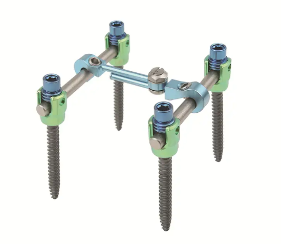
With all screws securely placed, the next phase involves connecting them with contoured rods. The rods must be shaped to match the patient’s anatomical or corrected spinal alignment. This step often requires specialized benders to achieve a smooth, accurate curvature. The rods are then seated into the screw heads. If spinal reduction is necessary, various reduction instruments are used to gently manipulate the spine, bringing it into alignment before the rods are fully secured. Finally, set screws are torqued down to lock the entire construct—screws and rods—into a single, rigid unit. This creates the stable spinal fixation system for thoracolumbar stabilization required for bone healing.
The procedure of inserting a spinal fixation device is a testament to surgical precision and advanced engineering working in concert. The tools provided must offer reliability and versatility to adapt to the unique demands of each case. At WEGO Medical, our contribution to this field is the Premier™ Thoracolumbar Posterior Internal Fixation System. Developed from over a decade of clinical experience, this Modular Spinal Fixation System is designed to offer spine surgeons versatile, safe, and efficient solutions for these complex procedures, supporting the critical work of restoring spinal stability.